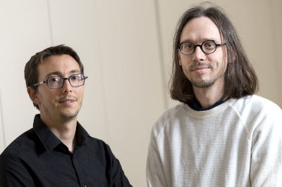
Co-team leaders : Harmen Reyngoudt & Benjamin Marty
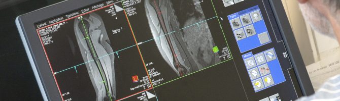
Nuclear magnetic resonance (NMR) imaging and spectroscopy are powerful techniques that can be used to study muscle anatomy, physiology and biochemistry during one single examination.
In addition, muscle intrinsic contrast with NMR is incomparably richer than with other techniques and optimized radiofrequency pulse sequences allow quantifying specifically different variables, like the intramuscular fat fraction, muscle water relaxation times, muscle perfusion and oxygenation, which can be altered in patients with neuromuscular disorders.
Our laboratory focuses most of its efforts in the development of these quantitative NMR methods, their validation, their promotion and their implementation in mono-centric and multi-centric clinical trials related to neuromuscular disorder. These trials allow studying both the natural history of the pathology or the effect of a novel therapeutic approach.
More precisely, our research projects aim at:
- Developing novel NMR contrasts in preclinical and clinical settings aiming at detecting and quantifying different pathophysiological processes involved in the onset and progression of myopathies, like muscle inflammation, necrosis and fibrosis.
- Determining the prognostic value of these novel NMR variables on disease progression in several neuromuscular disorders
- Developing tools to investigate the muscle at exercise (non-magnetic ergometers, multinuclear interleaved data acquisitions, …).
- Accelerating NMR data acquisition using novel acquisition schemes (undersampled radial encoding) and multi-parametric data acquisitions (MR-fingerprinting, deep learning approaches, …).
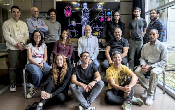
Team members
Benjamin Marty, PhD, Co-teamleader
Harmen Reyngoudt, PhD, Co-teamleader
Isabelle Ackermann-Bonan, MD, radiologist
Pierre-Yves Baudin, PhD, Design engineer
Inès Benziane, Design engineer
Edouard Berling, MD, PhD student
Jean-Marc Boisserie, MRI technologist
Sébastien Bougnaud, PhD, Research engineer
Ericky Caldas de Almeida Araújo, PhD, Research associate
Julien Dousseau, Animal technician
Mathias Duventru, MRI technologist
Yves Fromes, MD, PhD, Physician, DR1
Eric Giacomini, Design engineer
Valentin Henriet, PhD student
Sophie Jouan, MRI technologist
Hugo Lacombe, PhD student
Béatrice Matot, PhD, Research associate
Nacira Mendjel, PhD student
Lucie Ranno-Charrier, PhD student
Louis Rigler, PhD student
Constantin Slioussarenko, PhD, Research associate
Alexandra van Dongen, PhD student
Eléonore Vermeulen, PhD student
Contact
General informations: info_rmn@institut-myologie.org
Last publications
- Slioussarenko C, Lapert M, Baudin PY, Marty B. Upper-body free-breathing Magnetic Resonance Fingerprinting applied to the quantification of water T1 and fat fraction. Med Image Anal. 2025 Oct;105:103699. doi: 10.1016/j.media.2025.103699. Epub 2025 Jul 1. PMID: 40602206.
- Vermeulen E, Baudin PY, Lapert M, Marty B. Quantitative muscle water T2 mapping using RF phase-modulated 3D gradient echo imaging. Magn Reson Med. 2025 Sep;94(3):1103-1118. doi: 10.1002/mrm.30545. Epub 2025 May 6. PMID: 40326588.
- Rauh SS, Baudin PY, Stojkovic T, Birnbaum S, Decostre V, Zanfongnon RL, Fromes Y, Hooijmans MT, Strijkers GJ, Hogrel JY, Olivier S, Marty B, Reyngoudt H. Multi-parametric quantitative MRI of the lower limb muscles in a longitudinal study of limb-girdle muscular dystrophy R9. PLoS One. 2025 Apr 28;20(4):e0321463. doi: 10.1371/journal.pone.0321463. PMID: 40293996; PMCID: PMC12036925.
- Slioussarenko C, Baudin PY, Marty B. A steady-state MR fingerprinting sequence optimization framework applied to the fast 3D quantification of fat fraction and water T1 in the thigh muscles. Magn Reson Med. 2025 Jun;93(6):2623-2639. doi: 10.1002/mrm.30490. Epub 2025 Mar 4. PMID: 40033965; PMCID: PMC11971504
- Caldas de Almeida Araujo E, Barthélémy I, Fromes Y, Baudin PY, Blot S, Reyngoudt H, Marty B. Comprehensive quantitative magnetic resonance imaging assessment of skeletal muscle pathophysiology in golden retriever muscular dystrophy: Insights from multicomponent water T2 and extracellular volume fraction. NMR Biomed. 2025 Jan;38(1):e5278. doi: 10.1002/nbm.5278. Epub 2024 Oct 21. PMID: 39434514; PMCID: PMC11602680.
- Baudin PY, Balsiger F, Beck L, Boisserie JM, Jouan S, Marty B, Reyngoudt H, Scheidegger O. A Minimal Annotation Pipeline for Deep Learning Segmentation of Skeletal Muscles. NMR Biomed. 2025 Jul;38(7):e70066. doi: 10.1002/nbm.70066. PMID: 40390325; PMCID: PMC12089922.
- Lopez Kolkovsky AL, Matot B, Baudin PY, Caldas de Almeida Araujo E, Reyngoudt H, Marty B, Fromes Y. Multiparametric Aging Study Across Adulthood in the Leg Through Quantitative MR Imaging, 1H Spectroscopy, and 31P Spectroscopy at 3T. J Magn Reson Imaging. 2025 Jan;61(1):347-361. doi: 10.1002/jmri.29368. Epub 2024 Apr 9. PMID: 38593265.
- Bolano-Diaz CF, Reyngoudt H, Wilson IJ, James MK, Smith FE, Caldas de Almeida Araujo E, Gordish-Dressman H, Hilsden H, Rufibach LE, Wallace D, Ward L, Stramare R, Rampado A, Smith M, Boisserie JM, Le Louer J, Foster S, Peduto A, Sato N, Tamaru T, Sawyer AM, Day JW, Jones KJ, Walter MC, Stojkovic T, Mori-Yoshimura M, Mendell JR, Pegoraro E, Straub V, Blamire AM, Carlier P, Diaz-Manera J; Jain COS Consortium. Modeling of Dysferlinopathy (LGMDR2) Progression: A Longitudinal Fat Fraction Analysis. Neurol Genet. 2025 Jul 29;11(4):e200283. doi: 10.1212/NXG.0000000000200283. PMID: 40740171; PMCID: PMC12310143.
- Gerhalter T, Marty B, Gast LV, Roemer F, Baudin PY, Trollmann R, Uder M, Carlier PG, Nagel AM. Longitudinal Follow-Up of Patients With Duchenne Muscular Dystrophy Using Quantitative 23Na and 1H MRI. J Cachexia Sarcopenia Muscle. 2025 Apr;16(2):e13812. doi: 10.1002/jcsm.13812. PMID: 40254293; PMCID: PMC12009636.
- Reyngoudt H, Baudin PY, Caldas de Almeida Araújo E, Bachasson D, Boisserie JM, Mariampillai K, Annoussamy M, Allenbach Y, Hogrel JY, Carlier PG, Marty B, Benveniste O. Effect of sirolimus on muscle in inclusion body myositis observed with magnetic resonance imaging and spectroscopy. J Cachexia Sarcopenia Muscle. 2024 Jun;15(3):1108-1120. doi: 10.1002/jcsm.13451. Epub 2024 Apr 13. PMID: 38613252; PMCID: PMC11154752.
- Slioussarenko C, Baudin PY, Reyngoudt H, Marty B. Bi-component dictionary matching for MR fingerprinting for efficient quantification of fat fraction and water T1 in skeletal muscle. Magn Reson Med. 2024 Mar;91(3):1179-1189. doi: 10.1002/mrm.29901. Epub 2023 Oct 23. PMID: 37867467.
