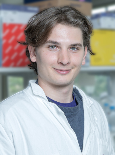
Dimitrios Kourtzas has been awarded the Master’s Prize for his research project on collagen VI (COLVI)-related disorders at the JSFM 2024. He just started a PhD under the supervision of Valérie Allamand, in the ‘Genetics and pathophysiology of neuromuscular diseases linked to the extracellular matrix and nucleus’ team led by Gisèle Bonne, at the Institute’s Center for Research in Myology. The PhD project is funded thanks to the DIM BioConvS (Région Ile de France). The Master prize was awarded to the young researcher in recognition of the quality of his Master work, and provides additional financial support for his thesis project, which is just getting underway. Interview with Dimitrios Kourtzas and Valérie Allamand.
What is the background to the work that has been rewarded?
Dimitrios Kourtzas: The prize relates to the work carried out in Valérie Allamand’s group as part of the 9-month internship I did for my Master’s degree at the University of Leiden in the Netherlands. The group’s theme deals with muscle diseases linked to collagen VI* for which no cure is available. These diseases are caused by a collagen VI deficiency, an extracellular matrix (ECM) protein present in most tissues.
My project was to finalize and validate, a model recreating an optimised pro-fibrotic environment in vitro, which we call “OPTIMEC”, and to use it for the first time to study diseases linked to collagen VI deficiency.
Valérie Allamand: The initial idea was to find optimal cell culture conditions for the expression and secretion of collagen VI. It was a project that I initiated at the University of Lund during my mobility (2019-2021), and which led to the OPTIMEC model for ‘optimised extracellular matrix’.
What results did you achieve?
DK: During my Master degree, I cultured skin fibroblasts from patients and controls in the OPTIMEC system to refine and validate the model. Creating a pro-fibrotic environment means that we want to reproduce the fibrosis process and therefore exacerbate the secretion of ECM components. We were able to verify this increase using various tools. We measured a significant increase in the quantity of collagen I, one of the best indicators of fibrosis, as well as collagen VI.
VA: Now we’re expanding the number of patient cells to make sure that it will be very useful for diagnosis, and to evaluate therapeutic strategies. Thanks to Myobank, Généthon and MyoLine, and our collaboration with numerous clinicians we have access to almost 300 cell cultures from patients with different mutations, which means we can be sure that the system is robust.
What are the possible applications of this optimized cell culture model?
DK: It can be used as a screening platform for anti-fibrotic therapies and in precision medicine approaches. But it will also be useful for understanding pathological mechanisms.
VA: Another reason for which we started to create this model, is to improve the diagnosis of collagen VI-related diseases. We hope to transfer our reliable model to diagnostic laboratories in the near future.
In addition, with such a robust and reproducible cell system, as Dimitrios mentioned, we can really test and validate pharmaceutical molecules or gene therapies and so select what is worth testing in greater depth in animal models.
What are the next steps?
VA: We are going to check that this system is compatible with cells derived from muscle tissue, and in particular fibroadipogenic progenitors (or FAPs), which are the main source of ECM components in muscle, and therefore of collagen VI.
This system will be useful for projects that will be conducted in collaboration with Capucine Trollet and Elisa Negroni (CdR team 3**, ANR funding), and with Juan Ma Fernandez-Costa (IBEC, Spain; AFM-Téléthon funding).
DK: My thesis, co-supervised by Valérie Allamand and Yves Fromes***, focuses on a gene therapy approach based on engineered tRNAs aimed at specifically correcting nonsense mutations that generate premature stop codons (PTCs), resulting in non-functional truncated proteins or lack of protein. This tool, developed by Christopher Ahern (Iowa University, USA) and his team, enables the synthesis of a protein that is not only full-length but also has an absolutely normal sequence. Thanks to the prize awarded by the SFM, we will be able to use cutting-edge proteomics techniques, in particular targeted proteomics to be able to detect very precisely the quantity of collagen VI that we will re-express.
VA: Dimitrios’ thesis is focused collagen VI deficiency, but as this tool is compatible with any gene, we will also be working on other forms of muscular dystrophy.
* Ullrich Congenital Muscular Dystrophy or Bethlem myopathy.
** Team 3: Cellular and molecular orchestration in muscle regeneration, in ageing and pathologies, led by Capucine Trollet and Vincent Mouly, at the Center of Research in Myology.
*** Yves Fromes works in the NMR and Spectroscopy Laboratory in the Neuromuscular Investigation Center at the Institute of Myology.
