Support our Foundation of Myology project

ALS: the thymus is both a target and a maintaining factor in the pathophysiology of the disease
Partager sur

Julia Pereira Lemos, a researcher in the Biotherapy for motor neuron diseases (ALS & SMA) in the Center for Research in Myology at the Institute, has just published an article* in the journal Brain Behav Immun Health on the role of the thymus in amyotrophic lateral sclerosis (ALS).
In what context did you conduct this research and what was the main objectives?

This research was conducted in the context of the growing recognition that ALS is not only a neurodegenerative disease but also involves complex interactions between the nervous and immune systems. Although regulatory T cells (Tregs) have been associated with neuroprotection and disease progression in ALS, it remained unclear whether some of these immune alterations could originate at the level of the thymus, where T cells differentiate and Tregs are generated.
Our main objective was to investigate whether ALS progression is associated with alterations in thymic structure and function, with a particular focus on T-cell development and the generation and function of Treg. More specifically, we aimed to characterize changes in the thymic microenvironment, thymocyte differentiation, and Treg development and function throughout disease progression.
It is particularly meaningful to see this work published, as it represents the result of a research project that we developed together, and I am very grateful to have had the opportunity to pursue this work within Piera’s group.
What methods did you use and what results did you obtain?
We used the SOD1-G93A transgenic mouse model of ALS, comparing mutant mice with age-matched wild-type controls at different stages of disease progression: 30 and 60 days (presymptomatic), 90 days (disease onset) and 120 days (late disease). We combined several complementary approaches, including flow cytometry, immunofluorescenceTechnique de visualisation d’une protéine spécifique sur des coupes tissulaires (réalisées à partir d’une biopsie) en utilisant un anticorps spécifique de cette protéine, sur lequel est fixé une substance fluorescente. and spatial analysis, western blotting, in situ hybridization, TREC quantification by qPCR, apoptosis assays, and in vitro« Dans le verre », expériences réalisées en laboratoire en dehors de l’organisme. Treg differentiation and suppression assays.
At the late stage of ALS, we observed marked thymic atrophy, with approximately 24% reduction in thymus relative weight and 40% reduction in total thymocyte numbers in male mSOD1 mice. There were also fewer double-positive thymocytes, CD4 single-positive cells, Treg progenitors and mature Tregs. Interestingly, Foxp3 expression itself was preserved, suggesting that the major problem was not simply loss of the Treg transcriptional program but a reduction in Treg generation and maintenance.
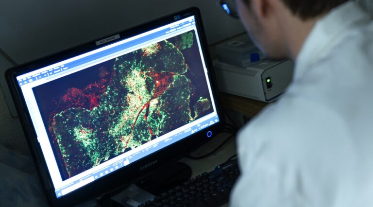
We also found important alterations in the thymic epithelial compartment, including reductions in cortical and medullary thymic epithelial cells and, particularly, mature medullary epithelial cells. In addition, Tregs showed abnormal localization, being found at greater distances from medullary epithelial cells and sometimes in cortical regions. This was accompanied by increased apoptosis and impaired Treg differentiation and suppressive function in vitro.
Finally, TREC analysis indicated alterations in thymocyte development, particularly around the transition toward the double-positive stage, while Treg numbers were also reduced in peripheral lymphoid tissues, especially in lymph nodes draining the affected hindlimbs.
What conclusions did you draw from this?
The main conclusion is that the thymus itself is affected during ALS progression and may represent an active component of ALS pathophysiology, rather than simply reflecting peripheral immune alterations.
Our results suggest that disease progression is associated with thymic atrophy, alterations of the thymic epithelial microenvironment, impaired thymocyte development, abnormal Treg localization, and reduced Treg differentiation and suppressive capacity. These changes could decrease the production and functional quality of immunoregulatory T cells and thereby contribute to the immune imbalance and neuroinflammation associated with ALS.
Overall, the data support a model in which the thymus participates in a bidirectional neuroimmune process: as ALS progresses, thymic dysfunction may contribute to peripheral immune dysregulation, which in turn may reinforce neuroinflammation and motor neuron degeneration. Importantly, this does not establish that thymic dysfunction is the initiating cause of ALS, but it identifies the thymus as a previously underappreciated component of the disease.
What are the perspectives for the future?
A major perspective is to investigate how ALS affects thymic function mechanistically. For example, our results raise questions about whether alterations in IL-2/TGF-β signalling, CD28 costimulation, extracellular matrix organization, integrin signalling, or the neuroimmune communication between the CNS and the thymus contribute to the observed defects. These mechanisms will require further experimental investigation.
From a translational perspective, the thymus could potentially become a target for approaches aimed at restoring Treg generation and function and re-establishing immune homeostasis. Future studies should determine whether restoring thymic/Treg function can influence neuroinflammation and disease progression, and whether markers of thymic function or immune signatures could help identify disease stages or patient subgroups.
More broadly, the perspective is not necessarily to replace existing ALS therapies, but to combine established neuroprotective treatments with approaches targeting immune dysregulation, guided where possible by biomarkers and immune gene signatures.
Andrew Ho : “This work provides enabling insights for the MRC project (GIANT), which starts from the premise that correcting a genetic defect does not by itself restore a healthy tissue, and that the host tissue is never a passive recipient of the therapy. By identifying the thymus as an organ actively compromised during ALS progression, with degraded epithelial support and impaired Treg generation and suppressive function, the study shows that immunoregulatory capacity is not constant but varies with disease stage. GIANT builds on this by integrating the immune context into the design of the therapeutic strategy, aiming for durable and effective transgene expression at reduced doses. “
** The project was initially conceived by Wilson Savino (a Brazilian researcher working at the Oswaldo Cruz Foundation (Rio de Janeiro), whose expertise in thymus biology and neuroimmune interactions provided important conceptual grounds for the study. I then had the opportunity to conduct this research within Dr. Piera Smeriglio’s group, where I developed the project and investigated the relationship between thymic function and immune dysregulation in ALS.