Support our Foundation of Myology project

Analysis of high-throughput digital images reveals distinct patterns of dystrophin expression in patients with dystrophinopathy
Partager sur
Duchenne muscular dystrophy (DMD) is the most common neuromuscular disease in boys while Becker’s muscular dystrophy (BMD), a milder and less progressive allelic variant, is much rarer. Both are recessive diseases linked to the X chromosomeForme que prend l’ADN pendant la division cellulaire (aspect de fins bâtonnets). Il est composé de 2 bras, un bras long et un bras court. Par convention, le bras long s’appelle q, et le bras court s’appelle p. Chez l’être humain, il y a 23 paires de chromosomes (soit 46 chromosomes). Vingt-deux paires sont constituées de 2 chromosomes identiques, appelés autosomes. La vingt-troisième paire est constituée des chromosomes sexuels, XX chez la femme et XY chez l’homme. with, in one case, a complete absence of dystrophin (DMD) and in the other, a partial deficiency of dystrophin (BMD). Both are linked to abnormalities in the DMD gene which encodes dystrophin, a protein whose quantity is closely related to the severity of the phenotype. Over the past decade, several antisense oligonucleotide drugs have been developed to induce the synthesis of a partially functional internally deleted dystrophin, similar to the one produced in BDM, and which improves the course of the disease. The mode of expression and functionality of dystrophin in patients with dystrophinopathy varies depending on several factors, such as its molecular functionality of dystrophin and its distribution. To assess the success of a therapeutic intervention, it is essential to understand the different modes of expression of dystrophin in these patients. Recently, several groups have used innovative techniques to quantify dystrophin in muscle biopsies from children but not in patients with a milder BMD.
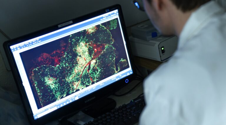
In this study, a team of European scientists involving researchers and clinicians from the Institute of Myology have been tackling dystrophin expression using both Western blotTransfert de protéines à partir d’un gel vers une membrane souple (nitrocellulose ou nylon) après séparation par électrophorèse. Cette technique est utilisée pour identifier une protéine. and an image analysis platform. automated high-throughput on skeletal muscle biopsies from DMD, BMD and DMD / BMD intermediates.
The results show a significant correlation between the Western blot and the immunofluorescenceTechnique de visualisation d’une protéine spécifique sur des coupes tissulaires (réalisées à partir d’une biopsie) en utilisant un anticorps spécifique de cette protéine, sur lequel est fixé une substance fluorescente. quantification, indicating the consistency between the different methodologies. They did, however, identify significant inter- and intra-disease heterogeneity in the expression patterns of dystrophin, regardless of the amount detected on the blot, due to the variability in fluorescence intensity and the sarcolemmal circumference coverage of dystrophin.
These results highlight the heterogeneity of the way dystrophin is expressed in BMD, which will help to evaluate dystrophin restorative therapies.