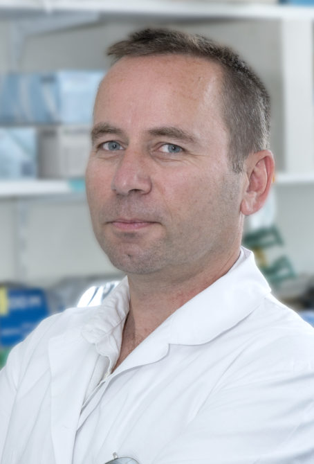
 Head: Bruno Cadot
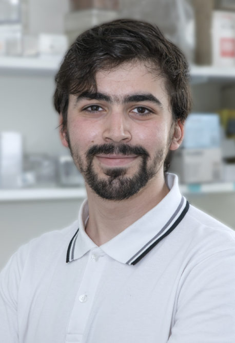
Assistant: Zoheir Guesmia
MyoImage develops myology-specific image analysis scripts using Open Source software such as FiJi, Python, Qupath and Cell Profiler, and supports users in the use of NIS, Zen and Metamorph software. In 2024, MyoImage met the needs of 9 teams for 100 image analysis projects, and carried out 2 external collaborative projects.
Head: Bruno Cadot
With a wealth of imaging experience acquired during his career, Bruno initiated the creation of the microscopy platform in 2017, of which he became the scientific manager. Over the past 7 years, he has developed the platform through acquisitions, improvements, user training and support, and the development of image analysis scripts.
Assistant: Zoheir Guesmia
After training in image analysis, medical imaging and electronics, Zoheir joined Myoimage as a design engineer in 2022. His skills enable him to meet the diverse analysis and support needs of researchers.
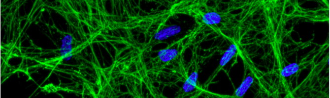
MyoImage has state-of-the-art imaging and image analysis equipment offering maximum imaging possibilities, while meeting quality requirements, via different approaches, from the infinitely small, with electron microscopy, to classic wide-field microscopy:
 • A Nikon Ti-2 confocal spinning microscope equipped with LiveSR, two cameras and an incubation chamber.
• A Nikon Ti wide-field microscope, equipped with an incubation chamber.
• NanoLive holotomography microscope, with incubation chamber.
• Zeiss wide-field microscope with Apotome module.
• Zeiss Axio Observer wide-field microscope.
• A Sartorius Incucyte for long-term monitoring of several culture plates.
• Two Life technologies Evos fluorescence microscopes.
• A Keyence VHX-S750 microscope for chemical staining.
• A Phillips CM120 electron microscope.
• Two image analysis workstations with AI-compatible graphics cards.
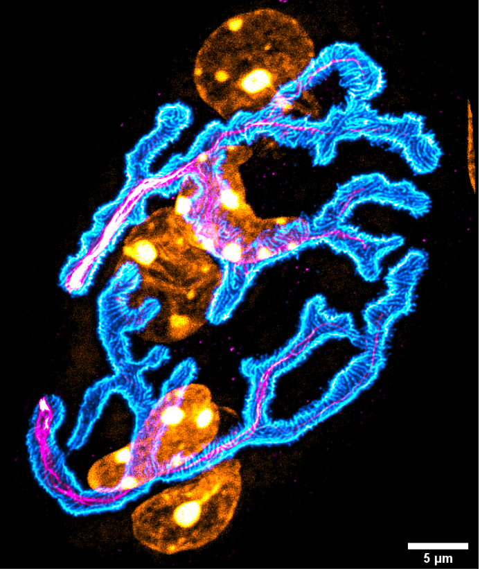
Neuromuscular Junction:
Nuclei (Orange), BTX (Cyan) and
Neurofilaments (Magenta).
Maximum projection of 43 z-slices
Key publications
- Traoré M, Noviello C, Vergnol A, Gentil C, Halliez M, Saillard L, Gelin M, Forand A, Lemaitre M, Guesmia Z, Cadot B, Caldas de Almeida Araujo E, Marty B, Mougenot N, Messéant J, Strochlic L, Sadoine J, Slimani L, Jolly A, De la Grange P, Hogrel JY, Pietri-Rouxel F, Falcone S. GDF5 as a rejuvenating treatment for age-related neuromuscular failure. Brain. 2024 Nov 4;147(11):3834-3848. doi: 10.1093/brain/awae107. Erratum in: Brain. 2025 Mar 6;148(3):e21. doi: 10.1093/brain/awae375. PMID: 38584513.
- Cardoso D, Guilbert S, Guigue P, Carabalona A, Harhouri K, Peccate C, Tournois J, Guesmia Z, Ferreira L, Bartoli C, Levy N, Colleaux L, Nissan X, Muchir A. Inhibition of poly(ADP-Ribosyl)ation reduced vascular smooth muscle cells loss and improves aortic disease in a mouse model of human accelerated aging syndrome. Cell Death Dis. 2024 Oct 2;15(10):723. doi: 10.1038/s41419-024-07078-7. PMID: 39353941; PMCID: PMC11448498.
- Wernert F, Moparthi SB, Pelletier F, Lainé J, Simons E, Moulay G, Rueda F, Jullien N, Benkhelifa-Ziyyat S, Papandréou MJ, Leterrier C, Vassilopoulos S. The actin-spectrin submembrane scaffold restricts endocytosis along proximal axons. Science. 2024 Aug 23;385(6711):eado2032. doi: 10.1126/science.ado2032. Epub 2024 Aug 23. PMID: 39172837.
