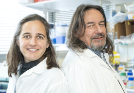
The results of an international study conducted by the Institute’s Capucine Trollet and Vincent Mouly team have just been published in Acta Neuropathologica. The article relates to the characterisation of PABPN1 aggregates performed on a unique series of 90 muscle biopsies from OPMD patients, collected from clinicians in France, Canada, the Netherlands and Israel, and on a xenograft model. This study is the culmination of a long-running project by the entire team, and more specifically by Fanny Roth, Jamila Dhiab and Alexis Boulinguiez. The project was initiated as part of the AFM eOPMD strategic project. Interview with Capucine Trollet.
What is the context of this work?
Oculopharyngeal muscular dystrophy (OPMD) is a rare muscle disease characterised by the appearance of pharynx and eyelid muscle weakness. The disease is caused by polyalanine expansion in the Poly (A) Binding Protein Nuclear 1 (PABPN1) protein, which causes the formation of intranuclear inclusions or aggregates in the muscles of patients with OPMD.
Despite a number of studies highlighting the harmful role of nuclear inclusions in cell and animal models of OPMD, their exact contribution to the human disease is still not clear.
What was this study’s objective and how did you proceed?
Our goal is to improve our understanding of the pathophysiology of OPMD. To this end, we performed an in-depth analysis of PABPN1 aggregates, according to age, genotype and muscle state. To this end, we used a significant and unique collection of human muscle biopsy samples.
Furthermore, in order to confirm what happens to PABPN1 aggregates in a regenerating muscle, we generated a xenograft model by transplanting human OPMD muscle biopsy samples into the hind legs of immunodeficient mice.
What results have you obtained?
We have shown, here, that age and genotype influence PABPN1 aggregates: the percentage of myonuclei containing PABPN1 aggregates increases with age and the HSP70 chaperone protein is more frequently co-located with aggregates that have larger polyalanine expansions.
In addition to the PRMT1 and HSP70 cofactors previously described, we have identified new components of the PABPN1 aggregates, in particular GRP78/BiP, RPL24 and p62.
We also observed that the myonuclei containing aggregates are larger than those not containing aggregates. In comparing two muscles from the same patient, a similar quantity of aggregates is observed in different muscles, except for the pharyngeal muscle, where less aggregates are observed. This could be due to the special nature of this muscle, which presents a low level of PABPN1 expression and which contains regenerating fibres.
Finally, the xenografts from subjects with OPMD showed regeneration of the human myofibres and the PABPN1 aggregates appeared quickly – in small quantities – after regeneration of the muscle fibres.
What conclusions did you draw?
Our data, obtained from human OPMD samples, support the non-exclusive models currently proposed for OPMD, which combine toxic PABPN1 intranuclear inclusions and a loss of PABPN1 function, which together result in this late-onset disease that selectively affects certain muscles.
* Team 3 – Cell and Molecular Orchestration in Muscle Regeneration, Ageing and Diseases at the Myology Centre for Research
