 Thomas Poulard* and Damien Bachasson** from the Neuromuscular Physiology and Evaluation Laboratory headed by Jean-Yves Hogrel, have just published, in the Journal of Physiology, the results of a study*** to develop an ultrafast ultrasound sequence making it possible to “film” diaphragm performance during cervical magnetic stimulation (CMS) of the phrenic nerves in healthy subjects.
What are the origins/overall context of this project?
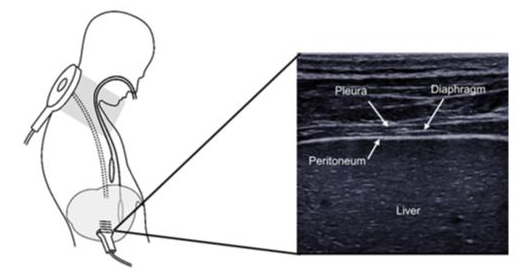
This work is part of the RespiMyo project, one of the objectives of which is to better understand diaphragm function using ultrasound. The function of the diaphragm can be defined as its ability to generate pressure, in particular allowing the exchange of gases in the lungs. Measuring the pressure generated by the diaphragm (transdiaphragm pressure (Pdi)) requires the use of oesophageal and gastric catheters. Pdi is then calculated as the difference between gastric pressure and oesophageal pressure. The measurement of Pdi is regularly associated with magnetic stimulation of the phrenic nerves (“Cervical Magnetic Stimulation” or CMS). The latter causes involuntary contraction of the diaphragm and this method is considered to be the reference technique in assessing diaphragm function. Coupling Pdi and CMS makes it possible to establish the “Pdi twitch” (Pditw), corresponding to the pressure generated by the diaphragm following CMS. However, measuring Pdi is invasive and using oesophageal and gastric catheters requires a high level of expertise. As a result, the use of ultrasound has been developed extensively in the last two decades, with the aim of observing diaphragm performance during tidal breathing. However, conventional ultrasound only allows a modest number of images per second (around 50) to be obtained. CMS-induced contraction lasts for a very short period of time, approximately 300 ms.
 What are the study objectives?
We therefore developed an ultrafast ultrasound sequence, making it possible to “film” diaphragm performance during CMS. Our goals were to: i) observe diaphragm performance with different CMS intensities, and ii) assess the link between ultrasound parameters and Pditw.
Which methods and protocols were used?
This study was conducted in healthy subjects. Thirteen participants were equipped with catheters allowing Pdi to be measured. We also developed an ultrafast ultrasound sequence allowing one ultrasound image to be obtained every millisecond. The participants were then subjected to a series of magnetic stimulations, at between 30% and 100% stimulator intensity. The ultrasound parameters that were studied were diaphragm tissue velocity (Vdi), and also diaphragm thickening fraction (TFdi). We then established the relationship between these ultrasound parameters and stimulation intensity, and also Pditw (i.e. the reference method).
What results were obtained?
We showed that there is a significant linear relationship between Vdi and stimulation intensity, for all participants. In other words, an increase in stimulations intensity is accompanied by an increase in Vdi. As for the relationship between TFdi and stimulation intensity, this was significant in 10 of the 13 participants. Our results also revealed a significant relationship between Vdi and Pditw in all participants. The relationship between TFdi and Vdi was significant in 8 of the 13 participants. Our data also show that the measurement of Vdi is very reliable, with reliability criteria close to those of Pditw.
What conclusions can be drawn and what opportunities has this study opened up?
This is the first study in which the diaphragm has been directly observed in response to CMS. We have shown that it is possible to extract ultrasound parameters that are reliable and sensitive to Pditw variations, in all participants. The application of this ultrafast ultrasound technique could be of great benefit in a population presenting diaphragm dysfunction (i.e. myopathy patients, patients under artificial ventilation, etc.). Indeed, the assessment of diaphragm function over time could be based on the measurement of Vdi, rather than Pditw, allowing the limitations associated with measuring Pdi to be overcome. The diagnostic power of this approach and its benefit in monitoring diaphragm dysfunction will be studied in patients in an upcoming study.
*Thomas Poulard, MSc, is a doctoral student at the Physiology and Neuromuscular Assessment Laboratory at the Institute of Myology (Paris, D. Bachasson, PT, PhD) and at the BioMaps Laboratory (University of Paris Saclay, J-L. Gennisson, PhD)
**Damien Bachasson PT, PhD, is a researcher at the Physiology and Neuromuscular Assessment Laboratory at the Institute of Myology (Paris)
***This study was carried out in collaboration with the BioMaps laboratory (Université Paris-Saclay ; Jean-Luc Gennisson), APHP-Sorbonne University R3S department (Martin Dres, Thomas Similowski), and the Relations between the nervous system and the respiratory system Laboratory (MRSU 1158 – Sorbonne University). This study is part of the RESPIMYO project supported by the EDF foundation.
