

Interview with Norma B. Romero and Stéphane Vassilopoulos
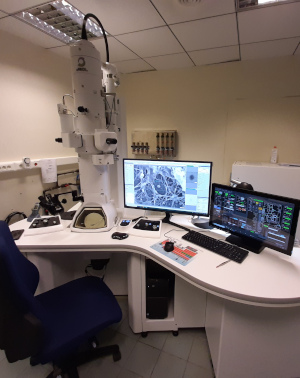
Since July 2021, researchers, doctors and engineers/ technicians from the Institute of Myology have been learning to use the different functions of the new state-of-the-art transmission electron microscope that the Institute has just acquired. While all the teams at the Myology Research Centre and the Institute’s Morphology Unit use electron microscopy, it is a crucial activity for the Norma B. Romero* and Stéphane Vassilopoulos** teams, both for research and diagnosis.
“The electron microscope that we have been using up to now, dates back to when the Institute of Myology was created, in 1996, and it has allowed us to make amazing discoveries;” notes N. B. Romero, “however, the device we have just acquired provides an image quality and level of detail and analysis that will allow us to continue our research and diagnostic work with even greater efficacy. ”
The cell structures, organelles, membranes, and also the abnormalities in these fine structures, are even more clearly visible, which allows our researchers to study to an even greater level of detail.
“We are extremely happy to be working with this new microscope, which is more powerful than the previous one, and thank Association Institut de Myologie and AFM-Téléthon for helping to fund it.” adds S. Vassilopoulos.
The electron microscope, an essential step in the process of understanding diseases
NBR. « In the last few years, there have been major advances in molecular genetics – a number of genes, mutations and myopathies have been identified – and knowledge of a muscular disease requires the ability to observe changes involved at the level of the muscle cells, organelles and proteins. It is crucial that we understand the pathophysiology of the cells and the disruption caused by the disease, so that we can then consider possible therapeutic solutions. »
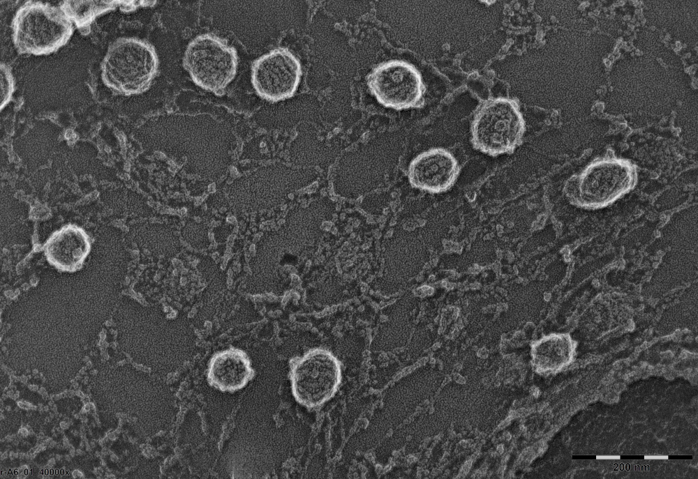
Fig. 1
SV. « In an era when molecular biological techniques are enabling therapies such as the ones we are developing at the Institute of Myology and at other institutes supported by AFM-Téléthon, it is essential that we are able to visualise the effects of these therapies inside our muscles at the molecular scale. It is essential that we are able to observe the beneficial effects of the treatments with photon microscopes, but also with this type of electron microscope, which is more powerful and higher-performing. It helps us to better visualise the diseased tissue and how a cure can be provided. »
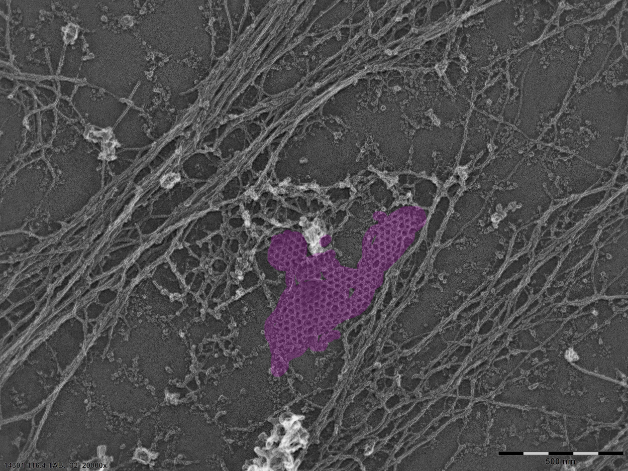
Fig. 2
Correlative microscopy, a specialty of the Institute
SV. « The Institute of Myology is one of the few laboratories in the world where correlative microscopy is being performed. To achieve this, we combine the techniques of super-resolution photon microscopy with electron microscopy techniques, and we superimpose the two images of the same cell. As such, we have, simultaneously, information about the structure with electron microscopy and information about protein composition or amounts under fluorescence; therefore, we have twice as much information about objects observed at the nanometre scale.
In the short term, it will be possible to see the proteins involved in the myopathies and understand how they are organised. For example, we have a project with Helge Amthor (University of Versailles) relating to the detection of dystrophin on the surface of muscle cells using fluorescence. For the moment, we do not see it at the ultrastructural level, but the use of the electron microscope will help us to understand how it interacts with the cytoskeleton of the muscle cells. »
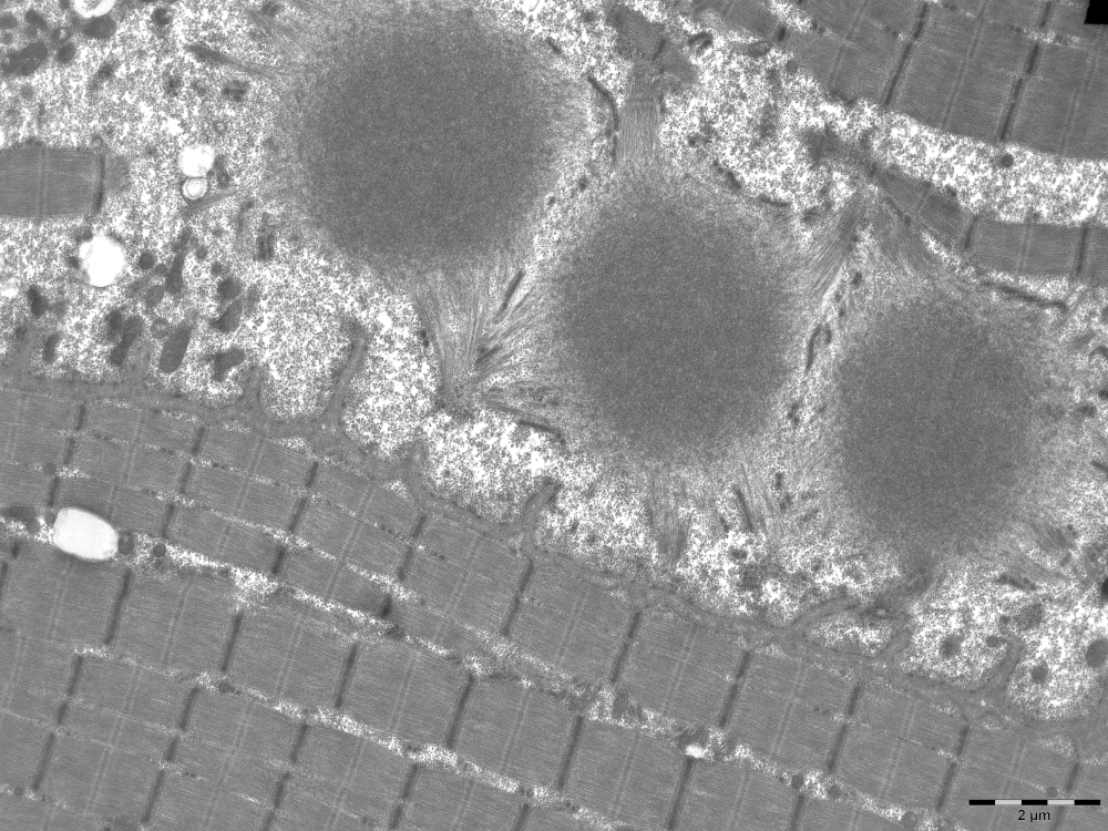
Fig. 3
Expertise that has been developed for years at the Institute of Myology
Electron microscopy is an expertise that has been developed at the Institute of Myology since its creation, resulting from the work by René Couteaux, Michel Fardeau and Fernando Tomé, global specialists in electron microscopy. This technique requires years of training, not only to operate the equipment, but also to understand what is being observed.
NBR. « Thanks to our cutting-edge equipment, we can discuss diagnoses or examine samples that are sent to our laboratory by external entities. For example, we are currently collaborating with a team that has studied cases of COVID-19 affecting the muscles, and that entrusts us with samples for advanced electron analysis. Furthermore, we have acquired a great deal of
experience in neonatal and congenital muscular disease, for which electron
microscopy analysis is crucial, essential. »
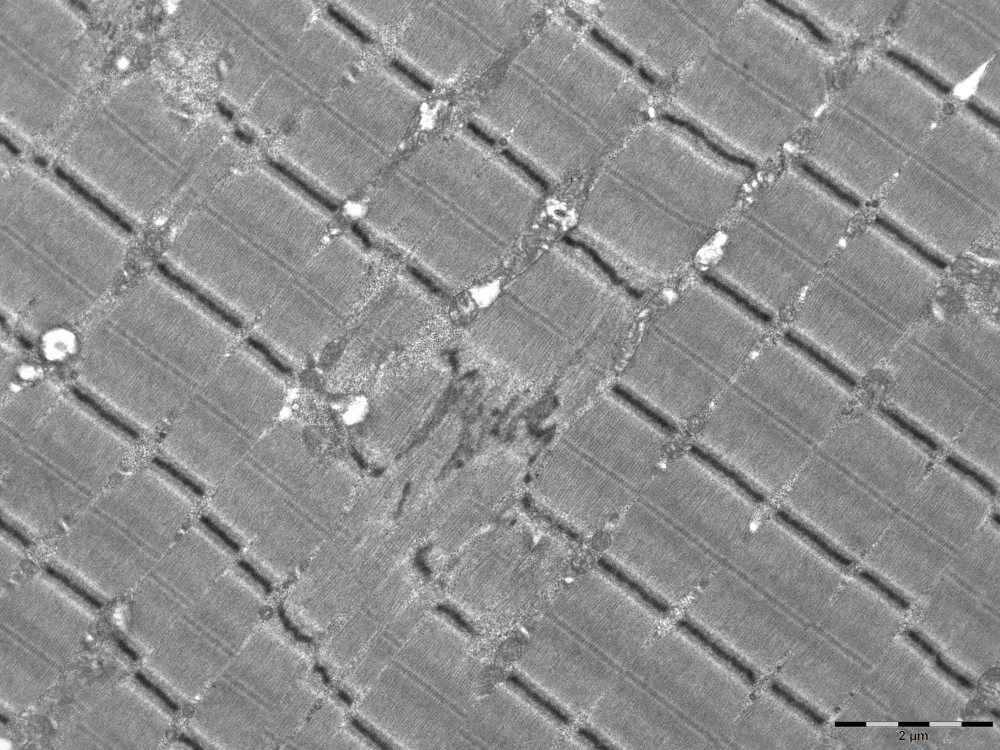
Fig. 4
SV. « We use microscopes that offer the highest imaging resolution, allowing us to perform a precise analysis of the diseased muscle. We have real expertise in the interpretation of this type of image. Indeed, researchers from laboratories all over the world consult us to obtain our opinion regarding samples or procedures. »
NBR. « I would also add that the electron microscopy photos that we take and that are identified, are also added to the Institute of Myology’s Muscle atlas, the first image database of the muscle, for well-characterised diseases, that we created with Bruno Cadot in September 2020. »
*Norma B. Romero, MD, PhD, is the head of the Morphological Unit at the Institute of Myology
** Stéphane Vassilopoulos, PhD, works in the Muscle Organization & Therapy of Dominant Centronuclear Myopathy team headed by Marc Bitoun in the Myology Centre for Research at the Institute of Myology
***The Achilles’ heel of dystrophin, large sarcolemmal protein, is its bonding with the cytoskeleton. It bonds with actin, the microtubules, and while we don’t know how it is anchored, we know that when this anchoring is defective, the muscle falls apart like a house of cards, because there is no longer this fastening component between the inside and outside of the muscle cell.
Fig.1. Analysis of the components of the membrane of muscle cells by transmission electron microscopy on replica platinum. This image shows structures called caveolae which are particularly abundant on the surface of muscle cells. In order to visualize the caveolae, the cultured muscle cells are scoured with ultrasound, they are fixed, quickly frozen and then cold dehydrated before the production of a metallic replica of their surface by rotary shading.
Fig.2. Analysis of the components of the membrane of muscle cells by transmission electron microscopy on replica platinum. Analysis of the components of the membrane of muscle cells by transmission electron microscopy on replica platinum. This image shows structures called caveolae which are particularly abundant on the surface of muscle cells. In order to visualize the caveolae, the cultured muscle cells are scoured with ultrasound, they are fixed, quickly frozen and then cold dehydrated before the production of a metallic replica of their surface by rotary shading.
Fig.3. Electron micrographs of skeletal muscle fibers in longitudinal sections showing three cytoplasmic bodies.
Fig.4. Electron micrographs of skeletal muscle fibers in longitudinal sections showing focal disorganization of the sarcomeric structure (congenital myopathies).
