The COVID-19 vaccination campaign is being discussed everywhere. It raises multiple questions, but do we know why vaccines, and in particular messenger RNA vaccines, have to be injected intramuscularly (into the muscle)? Here, we help you understand how the muscle optimises vaccination efficacy.
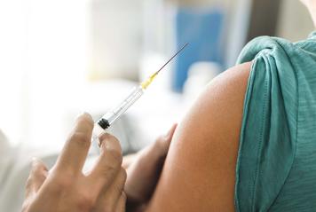
Whether it’s a vaccine for COVID-19, the flu or hepatitis B, the optimum route of administration is, for several different reasons, the same: an injection into the muscle, most commonly the shoulder muscle, the deltoid. If we go back to basics: in the context of a mass vaccination strategy, 4 key criteria must be respected: efficacy, safety, accessibility and reproducibility of the route of administration. There are 3 conventional routes of administration for vaccines: subcutaneous, intradermal or intramuscular. A consensus seems to have been established regarding the choice of method of administration, depending on the nature of the vaccine: the subcutaneous route is preferred for so-called “live” vaccines containing attenuated infectious agents (measles, mumps, rubella, etc), whereas the intramuscular route is favoured for so-called “inert” vaccines that no longer have an infectious agent present (the flu, COVID-19, Hepatitis B, etc.). Generally, immunogenicity – the ability to induce an immune response – and local tolerability – reaction at the injection site, oedema for example – are the same for both methods, but the muscle presents several significant advantages.
The muscle: a key role in immune response
Our muscles, which make up almost 50% of our body mass, have some well-kept secrets, notably the fact that they act as allies with our immune systems. Indeed, muscles release myokines, i.e. molecules that boost our immune defences and the fact that these muscles are highly vascularised means that the desired immune response is optimised.
During vaccination, the muscles activate several different mechanisms that produce antibodies. One of these mechanisms is based on the ability of muscle cells to express one or more proteins specific to the infectious agent. This protein behaves like an antigen, i.e. a molecule capable of being recognised by the immune system and activating the B and T cells, two types of white blood cell, that will come to recognise this antigen as a foreign substance and secrete antibodies to combat the infection.
In addition, the muscle is highly vascularised, i.e. it has numerous blood vessels, and so it also stimulates the macrophages, key immune system cells that “clean” the infected cells, then present the antigen to the B cells, causing them to produce antibodies, but also to the killer T cells, whose role it is to eliminate the cells recognised as being infected. Another way of calling on our immune system.
In the context of messenger RNA vaccines, the RNA acts as genetic instructions given to the cells into which it is injected. The RNA is translated by the ribosomes; these structures are present in the cells and synthesise the proteins by decoding the information contained in the RNA. They are found in particularly large quantities in the muscles. In practical terms, following the vaccination, the ribosomes spring into action and assemble, themselves, the component against which our body will learn to defend itself (in this case, a protein from the coronavirus viral envelope, the “spike” protein, for the COVID-19 vaccine). Because the muscle contains a significant amount of ribosomes, it acts as an optimal channel for the administration of a messenger RNA vaccine.
So, it should now be clear that we administer vaccines into the muscle to stimulate optimal antibody production. From the stapedius (in the ear), the smallest muscle in the human body, to the gluteus maximus, the largest, every muscle has a significant immune response capacity. We should consider ourselves lucky that, for reasons of accessibility and reproducibility, the specialists have now decided on the deltoid muscle (in the shoulder) for vaccine administration, and not the gluteus maximus, which was favoured 20 years ago, then abandoned in order to protect the sciatic nerve!
We would like to thank the researchers at the Institute of Myology, who helped to answer all these questions!
