PhD opportunity at the Nuclear Magnetic Resonance Laboratory at the Institute of Myology.
Low Field MR-Fingerprinting for respiratory assessment
Keywords: Low-field MRI, sequence development, undersampled reconstruction and optimization, AI-based denoising, respiratory function.
➣ Context
Pulmonary and muscle tissues involved in the respiratory function can be affected in various diseases (COPD, neuromuscular diseases, CoViD…). Quantitative MRI has become a key tool for studying neuromuscular diseases(1), but it mainly focuses on static limb muscles. Imaging the lungs and respiratory muscles is challenging due to motion and low MR signal levels. Current diagnostic tools have limitations, such as low sensitivity or radiation exposure. In this context, having access to tissue composition (fat fraction, vascularization…) on top of functional measures would be of great interest but is particularly challenging.
This PhD project aims at exploring jointly quantitative MRI and low-field MRI. Quantitative MRI provides valuable biomarkers, but requires long acquisition, while low-field MRI offers better accessibility, improved contrast and reduced field inhomogeneities(2), making it ideal for thoracic imaging. Nevertheless, the reduced signal-to-noise ratio is still a challenge in low-field MRI.
➣ Framework
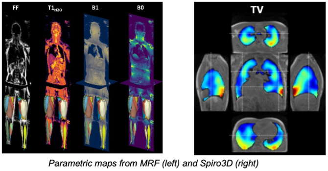
Magnetic Resonance Fingerprinting (MRF) is a new method(3) designed to overcome these challenges. By varying acquisition parameters, MRF generates a unique signal “fingerprint” for each tissue, enabling simultaneous extraction of multiple quantitative parameters through pattern recognition. Since MRF is less sensitive to noise, it is particularly promising for low-field MRI, potentially improving respiratory imaging(4). An MRF sequence(5), compensating the respiratory motion MoCo MRF T1-FF (6) has been designed and validated at the Institute of Myology for the characterization of muscle tissues by measuring 5 parameters simultaneously(7), on a 3T MRI (Siemens Healthineers). These developments will be the basis for a novel MRF sequence sensitive to additional parameters of interest (T2, water T2, vascularization) and adapted to the low-field constraints.
The validation on low field acquisitions will be done at the BioMaps laboratory, which recently acquired a 0.55 T MRI (Siemens Healthineers). To pave for the low SNR at 0.55 T, existing iterative reconstruction algorithms implemented at the Institute of Myology will be leveraged.
A 3D magnetic resonance spirometry sequence Spiro3D (8) was developed at the Biomaps laboratory providing a rich set of 3-dimensional parameters characterizing respiratory function and mechanics spatially. It was validated on healthy subjects and is currently being adapted to the 0.55 T scanner.
Both sequences are already running on clinical sites at 1.5T and 3T. The quantitative tissue measurements obtained by MRF will be compared against the functional measurements allowed by Spiro 3D on a small cohort of healthy subjects and patients.
➣ Objectives
The objective of this PhD project is to develop, optimize, and test 3D MR Fingerprinting techniques to quantify key parameters for the lungs and respiratory muscles. The PhD candidate will contribute to the following advancements:
- Development of MR Fingerprinting sequences sensitive to key respiratory parameters
- Optimization framework for MR Fingerprinting sequences under low-field constraints
using digital twins - Reconstruction of denoised 3D quantitative maps
- Validation of quantitative measurements against functional assessments in-vivo on healthy subjects and patients
➣ Environment
This PhD work will be split between the sites of BioMaps and Institute of Myology, in collaboration with Siemens Healthineers. It will be directed by Benjamin Marty (Co-head of the NMR laboratory, Institute of Myology), and co-supervised by Constantin Slioussarenko (Senior Researcher, Institute of Myology) and Angéline Nemeth (Associate Professor, Biomaps, Paris- Saclay University). The ongoing collaboration with Siemens on both sites will be leveraged, with a potential partnership on this specific project.
During the thesis, the PhD candidate will have access to two MRI systems installed inside the two laboratories: 0.55 T MRI (Biomaps) and 3T MRI (Institute of Myology). Collaborations are already planned with the thoracic radiology services of Raymond-Poincaré and Pitié- Salpêtrière AP-HP hospitals and their 1.5 T MRI.
The future PhD candidate will be in close interaction with current PhD candidates from the European V|LF-Spiro3D project led by BioMaps. This project brings together eleven partners (laboratories, hospitals, industrial companies) from four European countries for the development of 3D MRI lung function assessment. A collaboration with the AMT Center, University of Aberdeen, specialized in fingerprinting in very low-field MRI, is also being explored.
➣ Candidate Profile
- Engineering degree or Master’s (M2) specializing in applied mathematics, physics, medical imaging, or related fields.
- Excellent programming skills, particularly in Python. C++ programming experience is a plus.
- Ideally, experience in AI/ML (training/validation).
- Scientific curiosity, interest in experimentation.
- Strong proficiency in English (C1 level).
➣ Contact
Benjamin Marty: b.marty@institut-myologie.org
Angéline Nemeth: angeline.nemeth@universite-paris-saclay.fr
Constantin Slioussarenko: c.slioussarenko@institut-myologie.org
Bibliography
- (1) Marty, B.; Baudin, P. Y.; Reyngoudt, H.; Azzabou, N.; Caldas de Almeida Araújo, E.; Carlier, P. G.; Loureiro de Sousa, P. Simultaneous Muscle Water T2 and Fat Fraction Mapping Using Transverse Relaxometry with Stimulated Echo Compensation. NMR Biomed. 2016, 29 (4), 431–443. https://doi.org/10.1002/nbm.3459.
- (2) Sarracanie, M.; Salameh, N. Low-Field MRI: How Low Can We Go? A Fresh View on an Old Debate. Front. Phys. 2020, 8, 172. https://doi.org/10.3389/fphy.2020.00172.
- (3) Ma, D.; Gulani, V.; Seiberlich, N.; Liu, K.; Sunshine, J. L.; Duerk, J. L.; Griswold, M. A. Magnetic Resonance Fingerprinting. Nature 2013, 495 (7440), 187–192. https://doi.org/10.1038/nature11971.
- (4) Liu, Z.; Lugogo, N.; Agarwal, P.; Hamilton, J. Feasibility of Lung MR Fingerprinting at 0.55T Using a Deep Image Prior Reconstruction; Toronto, ON, Canada; p 2675. https://doi.org/10.58530/2024/2675.
- (5) Slioussarenko, C.; Baudin, P.; Marty, B. A Steady‐state MR Fingerprinting Sequence Optimization Framework Applied to the Fast 3D Quantification of Fat Fraction and Water T1 in the Thigh Muscles. Magn. Reson. Med. 2025, mrm.30490. https://doi.org/10.1002/mrm.30490.
- (6) Slioussarenko, C.; Baudin, P.-Y.; Lapert, M.; Marty, B. Upper-Body Free-Breathing Magnetic Resonance Fingerprinting Applied to the Quantification of Water T1 and Fat Fraction. arXiv September 24, 2024. http://arxiv.org/abs/2409.16200 (accessed 2024-09-25).
- (7) Slioussarenko, C. Whole-Body Quantitative Imaging of the Skeletal Muscle by Magnetic Resonance Fingerprinting. Theses, Université Paris-Saclay, 2024. https://theses.hal.science/tel-04939151.
- (8) Barrau, N. 3D MR Spirometry. Theses, Université Paris-Saclay, 2024. https://theses.hal.science/tel- 04663088.
Download the complete announcement of the PhD opportunity at the Nuclear Magnetic Resonance Laboratory at Institute of Myology : Low Field MR-Fingerprinting for respiratory assessment