PhD opportunity at the Nuclear Magnetic Resonance Laboratory at Institute of Myology.
Development of real-time quantitative MRI for muscles involved in respiration, swallowing and eye movements
Keywords: Muscle MRI, real-time MRI, undersampled reconstruction, non-Cartesian MRI acquisition
➣ Context
Quantitative MRI (qMRI) is a well-established tool for the in vivo characterization and longitudinal monitoring of skeletal muscle pathology in neuromuscular diseases(1). It is increasingly used in clinical trials, particularly targeting appendicular muscles (limbs) through static imaging protocols such as T1-weighted, Dixon, and T2 mapping(2).
However, these conventional methods are limited in their ability to assess small, continuously active muscles involved in essential functions like respiration, swallowing, and eye movement—muscles that are often impaired in neuromuscular conditions and play a significant role in patient morbidity and mortality(3,4). These muscle groups remain underexplored due to challenges in imaging dynamic motion and achieving sufficient spatial resolution.
To address this gap, real-time MRI has emerged as a promising solution, enabling high- temporal-resolution imaging via rapid, undersampled acquisitions(5). Reconstruction techniques leverage either compressed sensing with temporal-spatial regularization(6) or deep learning approaches, which improve reconstruction fidelity and speed(7,8).
Originally applied in domains like cardiac cine imaging(9), real-time MRI offers a valuable platform for capturing the functional dynamics of non-limb muscles, with potential to deliver novel biomarkers for both disease progression and therapeutic efficacy assessment.
➣ Framework
A 3D radial MRI sequence (MoCo MRF T1-FF(10)) compatible with real-time imaging, along with its reconstruction pipeline, has been implemented at the NMR laboratory of the Institute of Myology and validated on a 3T MRI system (Siemens Healthineers). This development will serve as the foundation for a new real-time MRI sequence targeting respiratory muscles. In parallel, quantitative MRI protocols have been established to assess fat fraction and inflammation in both respiratory(10) and tongue muscles(11), enabling correlation between structural tissue biomarkers and functional metrics from real-time imaging. Additionally, in- house segmentation algorithms (museg-ai) could be adapted for dynamic tracking of muscles in motion. Functional assessments, such as tongue strength measurements, have been developed in collaboration with the laboratory of physiology and neuromuscular evaluation at the Institute of Myology, providing complementary data for validating MRI- derived functional biomarkers. 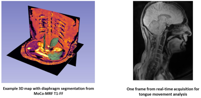
➣ Objectives
The objective of this thesis is to develop and optimize real-time MRI techniques in the context of quantitative imaging of dynamic skeletal muscles. The doctoral candidate will contribute to the following advancements:
- Real-time imaging and tracking of dynamic processes: Optimize acquisition sequences to capture and analyze the dynamics of muscles involved in respiration, swallowing, and eye movements.
- Optimization of 3D reconstructions: Advanced iterative reconstruction algorithms taking into account motion.
- Adaptation of segmentation algorithms for dynamic tracking of moving structures
- Development of functional biomarkers: Identify and quantify new biomarkers describing these processes to improve the characterization of functional alterations related to neuromuscular pathologies.
➣ Environment
This PhD work will be carried at the Institute of Myology. It will be directed by Benjamin Marty (Co-head of the NMR laboratory, Institute of Myology), and co-supervised by Constantin Slioussarenko (Senior Researcher, Institute of Myology). The ongoing collaboration with Siemens Healthineers will be leveraged, with one on-site clinical scientist.
During the thesis, the PhD candidate will have access to the 3T MRI system (PrismaFit Siemens Healthineers) installed at Institute of Myology. Existing collaborations with the radiology services of Raymond-Poincaré and Pitié-Salpêtrière AP-HP hospitals and their 1.5 T MRI could be leveraged as well for clinical applications. Biomechanical modelling with inputs from specialist laboratories are potential routes of investigation for developing biomarkers linked to the physiology.
The future PhD candidate will be in close interaction with current PhD candidates at Institute of Myology working on quantitative MRI and deep-learning muscles segmentation.
The funding for this PhD project is derived from the GenoTher biocluster project, part of the French Innovation Santé 2030 plan.
➣ Candidate Profile
- Engineering degree or Master’s (M2) specializing in applied mathematics, physics, medical imaging, or related fields.
- Excellent programming skills, particularly in Python. C++ programming experience is a plus.
- Ideally, experience in AI/ML (training/validation).
- Scientific curiosity, interest in experimentation.
- Strong proficiency in English (C1 level).
➣ Contact
Benjamin Marty: b.marty@institut-myologie.org
Constantin Slioussarenko: c.slioussarenko@institut-myologie.org
Bibliography
- (1) Marty, B.; Baudin, P. Y.; Reyngoudt, H.; Azzabou, N.; Caldas de Almeida Araújo, E.; Carlier, P. G.; Loureiro de Sousa, P. Simultaneous Muscle Water T2 and Fat Fraction Mapping Using Transverse Relaxometry with Stimulated Echo Compensation. NMR Biomed. 2016, 29 (4), 431–443. https://doi.org/10.1002/nbm.3459.
- (2) Carlier, P. G.; Marty, B.; Scheidegger, O.; Loureiro de Sousa, P.; Baudin, P.-Y.; Snezhko, E.; Vlodavets, D. Skeletal Muscle Quantitative Nuclear Magnetic Resonance Imaging and Spectroscopy as an Outcome Measure for Clinical Trials. J. Neuromuscul. Dis. 2016, 3 (1), 1–28. https://doi.org/10.3233/JND-160145.
- (3) Bourke, S. C. Respiratory Involvement in Neuromuscular Disease. Clin. Med. 2014, 14 (1), 72–75. https://doi.org/10.7861/clinmedicine.14-1-72.
- (4) Argov, Z.; De Visser, M. Dysphagia in Adult Myopathies. Neuromuscul. Disord. 2021, 31 (1), 5–20. https://doi.org/10.1016/j.nmd.2020.11.001.
- (5) Uecker, M.; Zhang, S.; Voit, D.; Karaus, A.; Merboldt, K.; Frahm, J. Real‐time MRI at a Resolution of 20 Ms. NMR Biomed. 2010, 23 (8), 986–994. https://doi.org/10.1002/nbm.1585.
- (6) Feng, L.; Axel, L.; Chandarana, H.; Block, K. T.; Sodickson, D. K.; Otazo, R. XD-GRASP: Golden-Angle Radial MRI with Reconstruction of Extra Motion-State Dimensions Using Compressed Sensing. Magn. Reson. Med. 2016, 75 (2), 775–788. https://doi.org/10.1002/mrm.25665.
- (7) He, Z.; Zhu, Y.-N.; Chen, Y.; Chen, Y.; He, Y.; Sun, Y.; Wang, T.; Zhang, C.; Sun, B.; Yan, F.; Zhang, X.; Sun, Q.-F.; Yang, G.-Z.; Feng, Y. A Deep Unrolled Neural Network for Real-Time MRI-Guided Brain Intervention. Nat. Commun. 2023, 14 (1), 8257. https://doi.org/10.1038/s41467-023-43966-w.
- (8) Ahmad, R.; Bouman, C. A.; Buzzard, G. T.; Chan, S.; Liu, S.; Reehorst, E. T.; Schniter, P. Plug-and-Play Methods for Magnetic Resonance Imaging: Using Denoisers for Image Recovery. IEEE Signal Process. Mag. 2020, 37 (1), 105–116. https://doi.org/10.1109/MSP.2019.2949470.
- (9) Longère, B.; Abassebay, N.; Gkizas, C.; Hennicaux, J.; Simeone, A.; Rodriguez Musso, A.; Carpentier, P.; Coisne, A.; Pang, J.; Schmidt, M.; Toupin, S.; Montaigne, D.; Pontana, F. A New Compressed Sensing Cine Cardiac MRI Sequence with Free-Breathing Real-Time Acquisition and Fully Automated Motion-Correction: A Comprehensive Evaluation. Diagn. Interv. Imaging 2023, 104 (11), 538–546. https://doi.org/10.1016/j.diii.2023.06.005.
- (10) Slioussarenko, C.; Baudin, P.-Y.; Lapert, M.; Marty, B. Upper-Body Free-Breathing Magnetic Resonance Fingerprinting Applied to the Quantification of Water T1 and Fat Fraction. arXiv September 24, 2024. http://arxiv.org/abs/2409.16200 (accessed 2024-09-25).
- (11) Vermeulen, E.; Baudin, P.-Y.; Lapert, M.; Marty, B. Quantitative Assessment of Tongue Tissue Structure with 3D Partially Spoiled Gradient Echo; Toronto, ON, Canada; p 0902. https://doi.org/10.58530/2024/0902.
Download the complete announcement of the PhD opportunity at the Nuclear Magnetic Resonance Laboratory at Institute of Myology: Development of real-time quantitative MRI for muscles involved in respiration, swallowing and eye movements